


 739 / 912

Chapter 21 731
Copyright © 2017 Pearson Education, Inc.
78.
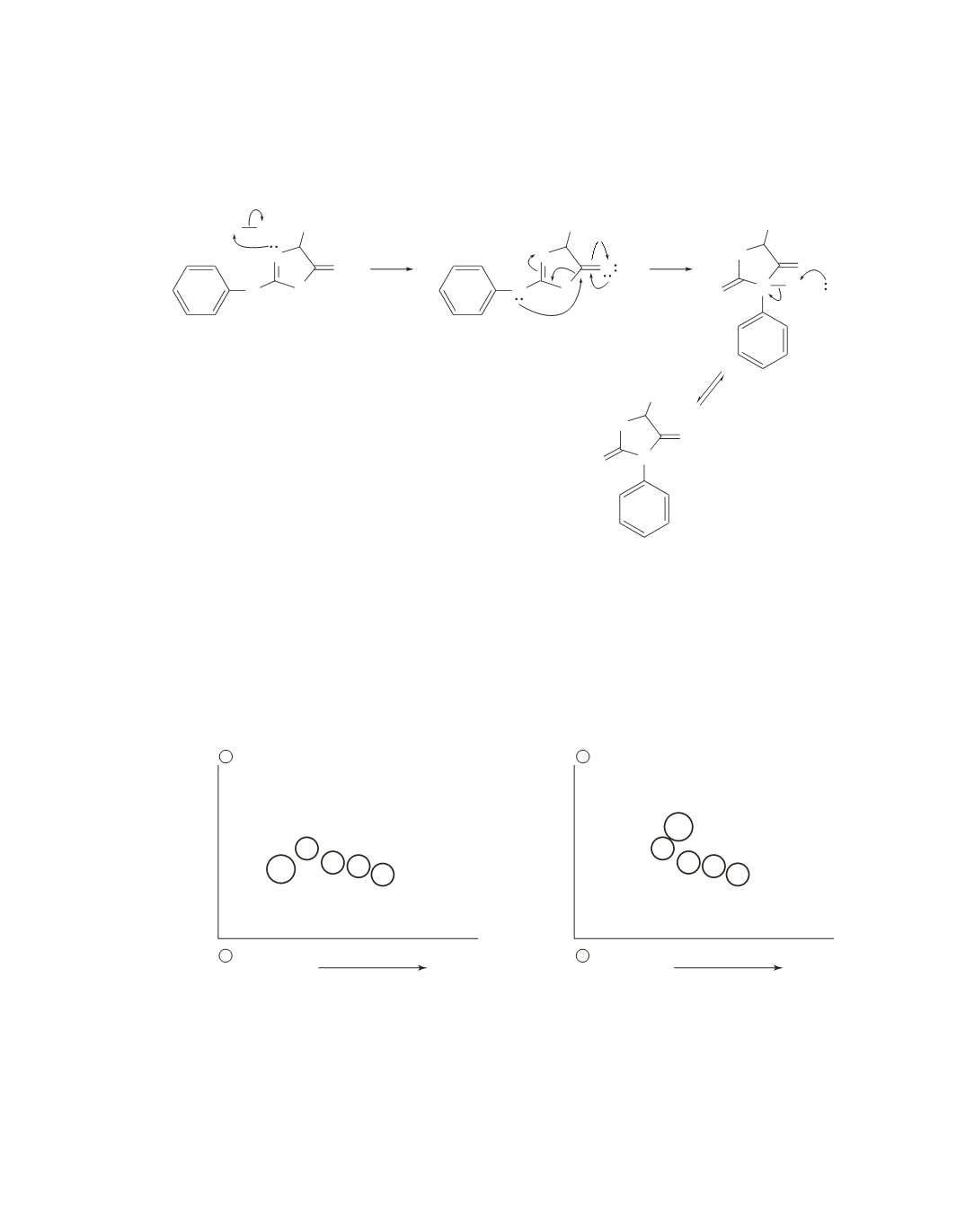
The acid protonates the lone pair that are
sp
2
electrons. Formation of the tetrahedral intermediate (the
two arrows that represent electron flow to the right) and collapse of the tetrahedral intermediate (the three
arrows that represent electron flow to the left) are shown together in the second step.
N
N
S
R
O
O
H
H
+
+
N
N
O
R
H
a thiazoline
a PTH-amino acid
N
N
R
H B
H
S
S
+
N
N
S
R
O
BH
H
79.
The spot marked with an
X
is the peptide that is different in the normal and mutant polypeptide. The spot
is closer to the cathode and farther to the right, indicating that the substituted amino acid in the mutant has
a greater pI and is less polar.
The fingerprints are those of hemoglobin (normal) and sickle-cell hemoglobin (mutant). In sickle-cell
hemoglobin, a glutamate in the normal polypeptide has been substituted with a valine. This agrees with
our observation that the substituted amino acid is less negative and more nonpolar. (See the discussion of
sickle-cell anemia on page 1171 of the text.)
−
+
−
+
X
X
paper chromatography
normal
mutant
electrophoresis at pH = 6.5
electrophoresis at pH = 6.5
paper chromatography